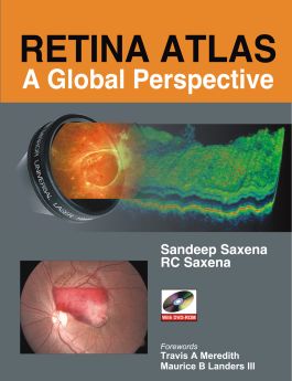
Retina Atlas: A Global Perspective
Receive via shipping:
- Print bound version of the complete text
1. Fluorescein Angiography and Indocyanine Green Angiography 3
2. Optical Coherence Tomography 15
3. B-scan Ultrasonography 23
4. Microperimetry 27
5. Multifocal Electroretinogram 31
6. Retina Viewing Lenses and Systems 39
SECTION 2
Part 1: Retinal Vascular Disorders
7. Diabetic Retinopathy 49
8. Central Retinal Vein Occlusion 129
9. Branch Retinal Vein Occlusion 147
10. Retinal Artery Occlusion 167
11. Plasma Protein Risk Factors for Retinal Vascular Occlusive Diseases 179
12. Ocular Ischemic Syndrome 183
13. Retinal Artery Macroaneurysm 187
14. Parafoveal Telangiectasis 193
15. Hypertensive Fundus Changes 209
16. Coats’ Disease 213
17. Eales’ Disease 219
18. Hemoglobinopathies 233
19. Retinal Vascular Changes Caused by Hematologic Disorders 237
20. Malarial Retinopathy 245
21. Dengue Hemorrhagic Fever 249
22. Takayasu’s Disease 253
23. Retinopathy of Prematurity 257
Part 2: Macular Disorders
24. Age-related Macular Degeneration 263
25. Angioid Streaks 341
26. Ocular Histoplasmosis 349
27. Central Serous Chorioretinopathy 353
28. Postsurgical Cystoid Macular Edema 383
29. Myopia 389
30. Photic Retinopathy 409
Part 3: Vitreoretinal Surgical Disorders
31. Epiretinal Membranes, Pseudoholes and Vitreomacular Traction Syndrome 413
32. Idiopathic Macular Hole 433
33. Retinal Detachment 455
34. Coloboma of the Choroid 477
Part 4: Inflammatory Disorders
35. Acute Posterior Multifocal Placoid Pigment Epitheliopathy 485
36. Serpiginous Choroiditis 493
37. Ampigenous Choroiditis 501
38. Multifocal Choroiditis 505
39. Punctate Inner Choroidopathy 515
40. Multiple Evanescent White Dot Syndrome 521
41. Birdshot Retinochoroidopathy 525
42. Sympathetic Ophthalmia 531
43. Vogt-Koyanagi-Harada Syndrome 539
44. Intermediate Uveitis 555
45. Subretinal Fibrosis and Uveitis Syndrome 563
46. Sarcoidosis 571
47. Behçet’s Disease 577
48. Systemic Lupus Erythematosus 581
49. Posterior Scleritis 585
Part 5: Infectious Disorders
50. Toxoplasmosis 589
51. Toxocariasis 599
52. Ocular Tuberculosis 603
53. Ocular Cysticercosis 613
54. Cat-scratch Disease 617
55. Leptospirosis 623
56. Fungal Diseases 627
57. Retinal and Optic Nerve Involvement in Syphilis 633
58. Autoimmune Deficiency Disease 637
Part 6: Traumatic Disorders
59. Posterior Segment Trauma and Choroidal Folds 651
60. Choroidal Rupture 657
61. Commotio Retinae 663
62. Valsalva Retinopathy 667
63. Purtscher’s Retinopathy 671
64. Subhyaloid Hemorrhage, Traumatic Macular Hole and Electric Burn 675
65. Retained Intraocular Foreign Body 683
66. Traumatic Optic Pit Maculopathy 689
67. Optic Nerve Evulsion and Hemorrhagic Choroidal Detachment 693
Part 7: Hereditary Chorioretinal Disorders
68. Retinitis Pigmentosa and Allied Disorders 699
69. Best’s Disease 729
70. Congenital X-Linked Retinoschisis 749
71. Progressive Cone Dystrophy and Cone-Rod Dystrophy 765
72. Pattern Dystrophy of the Retinal Pigment Epithelium 781
73. Stargardt’s Disease and Fundus Flavimaculatus 787
74. Congenital Stationary Night Blindness and Benign Fleck Retina Syndrome 807
75. Central Areolar Choroidal Dystrophy and North Carolina Macular Dystrophy 819
76. Choroideremia 825
77. Gyrate Atropy 833
78. Malattia Leventinese or Doyne Honeycomb Retinal Dystrophy 837
79. Bietti’s Crystalline Dystrophy 851
80. Albinism 859
81. Drug-induced Retinal Toxicities 865
Part 8: Tumors
82. Choroidal Nevus 891
83. Choroidal Melanoma 895
84. Choroidal Metastasis 901
85. Choroidal Hemangioma 905
86. Choroidal Osteoma 911
87. Capillary Hemangioma 915
88. Cavernous Hemangioma and
Racemose Hemangioma 925
89. Vasoproliferative Tumors of the Retina 929
90. Congenital Hypertrophy of the
Retinal Pigment Epithelium 935
91. Congenital Simple Hamartoma of
the Retinal Pigment Epithelium and Congenital Albinotic Retinal Pigment Epithelium Nevi 939
92. Combined Hamartoma of the Retina
and Retinal Pigment Epithelium 943
93. Astryocytic Hamartoma of the Retina 947
94. Retinoblastoma 951
95. Intraocular Medulloepithelioma 965
Part 9: Optic Nerve Disorders
96. Congenital Optic Disc Pit 969
97. Melanocytoma of the Optic Nerve 987
98. Optic Disc Drusen 993
Part 10: Miscellaneous
99. Retinal Diseases in Pregnancy 997
100. Cancer Therapy-associated
Retinopathy 1001
101. Myelinated Retinal Nerve Fibers 1009
102. Synchisis Scintillans and Asteroid
Hyalosis 1013
Index 1017
82. Choroidal Nevus 891
83. Choroidal Melanoma 895
84. Choroidal Metastasis 901
85. Choroidal Hemangioma 905
86. Choroidal Osteoma 911
87. Capillary Hemangioma 915
88. Cavernous Hemangioma and Racemose Hemangioma 925
89. Vasoproliferative Tumors of the Retina 929
90. Congenital Hypertrophy of the Retinal Pigment Epithelium 935
91. Congenital Simple Hamartoma of the Retinal Pigment Epithelium and Congenital Albinotic Retinal Pigment Epithelium Nevi 939
92. Combined Hamartoma of the Retina and Retinal Pigment Epithelium 943
93. Astryocytic Hamartoma of the Retina 947
94. Retinoblastoma 951
95. Intraocular Medulloepithelioma 965
Part 9: Optic Nerve Disorders
96. Congenital Optic Disc Pit 969
97. Melanocytoma of the Optic Nerve 987
98. Optic Disc Drusen 993
Part 10: Miscellaneous
99. Retinal Diseases in Pregnancy 997
100. Cancer Therapy-associated Retinopathy 1001
101. Myelinated Retinal Nerve Fibers 1009
102. Synchisis Scintillans and Asteroid Hyalosis 1013
Index 1017
Publisher's Note: Products purchased from Third Party sellers are not guaranteed by the publisher for quality, authenticity, or access to any online entitlements included with the product.
A lavishly-illustrated step-by-step guide to retinal disorders
This atlas, by two of the world's leading authorities, is extensively illustrated with hundreds of full-color clinical photographs. It delivers the step-by-step visual guidance on a wide range of retinal disorders, accompanied by differential diagnoses in side-by-side page layouts to assist the reader in identifying a full range of retinal disorders. It includes the basics of fluorescein angiography, idocyanine green angiography, scanning laser ophthalmoscope based angiography, time-domain and spectral domain high-resolution optical coherence tomography with perimeter and multifocal electroretinography.
This extremely timely, thorough, and well integrated book will be valuable to the active vitreoretinal specialist as well as comprehensive ophthalmologist.
Features:
- Global perspective
- More than 100 chapters
- DVD with spectral-domain high-resolution optical coherence tomography videos
- 2300 full-color images
- Step-by-step macular surgeries
- First retina atlas presenting the medical and surgical aspects of vitreoretinal diseases in a comprehensive manner

